
Figure 7. Molecular modeling of the interaction of LMF with the [(HSA-PPIX)-TF] complex, (A) and the second site of the interaction of LMF with [(HSA-PPIX)-TF] (B), represented as a solid ribbon, colored by secondary structure, LMF represented as sticks.
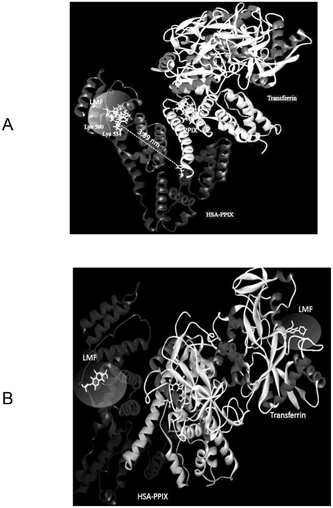
The docking position of LMF to the protein is highlighted. LMF was docked in sub-domain IIIB of (HSA-PPIX). The distance between the binding site candidates of LMF to Trp is also illustrated. The hydrogen bonds between LMF and (HSA-PPIX) are represented as green dashed lines.