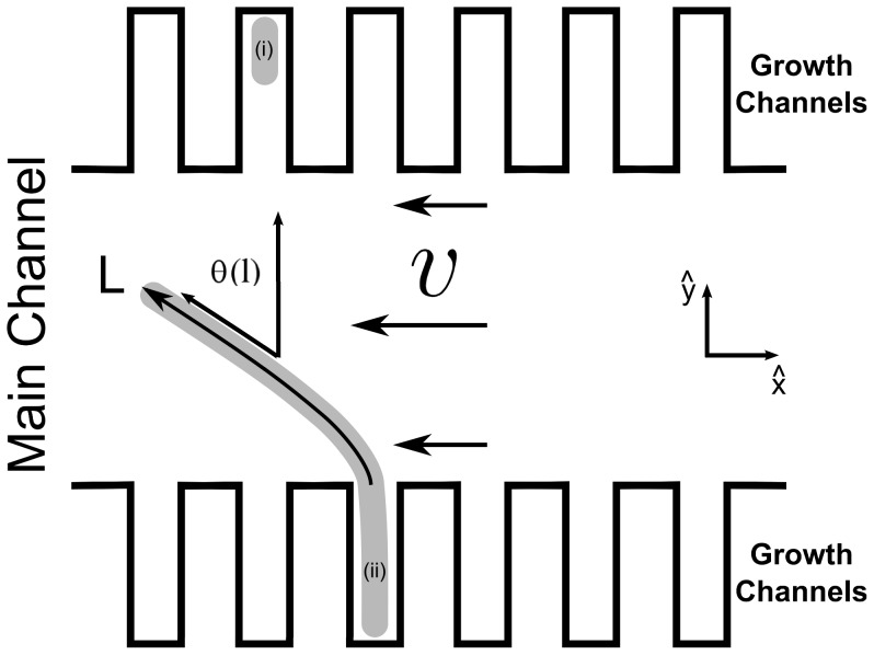
Figure 1. Sketch of the experimental setup.
Cells, e.g. cell (i) and (ii), grew in a microfluidic device consisting of dead-end growth channels that were connected at their open end to a large main channel. If cell division is blocked, the cells grow as filaments that penetrate into the main channel. A fluid flow with velocity  and a profile similar to the one depicted created a hydrodynamic force that deformed the cells, as is shown for cell (ii). The magnitude of the force was controlled by the infusion rate. For analysis, the arc length (
 ) was measured from the point of connection between the growth channels and the main channel (
 ) to the tip of the cell (
 ). To characterize the shape of the cell, the angle profile (
 ) was calculated (see Materials and Methods).

