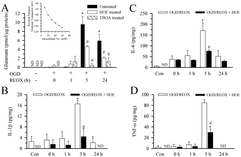
Figure 4. Glutamate and pro-inflammatory cytokine release from hippocampal astrocytes following OGD/REOX.
A. Glutamate release in hippocampal astrocyte cultures was determined at 2(1 µM) or TBOA (100 µM) was present during REOX only. Data are mean ± SEM. N of 4 cultures were for all groups except for normoxia (n = 5). *p<0.05 vs. normoxic control, # p<0.05 vs. corresponding untreated. ND: not detectable. Inset: The reversal potential for the glutamate transport was plotted as a function of [Na+]i. The known values for [Na]o, [H]o, [H]i and [K]o at baselines were used along with an assumed value for [Glu]o of 0.01 µM, [Glu]i of 5 mM and [K]i of 70 mM. B–D. Release of innate immune cytokines in the culture medium of hippocampal astrocytes. HOE 642 (1 µM) was present during normoxia or REOX treatment. IL-1β (B) IL-6 (C), or TNF-α (D) were normalized to cell lysate protein and expressed as pg/mg protein. Data are mean ± SEM (n = 3). ND: not detectable. *p<0.05 vs. normoxic control. # p<0.05 vs. corresponding untreated.

