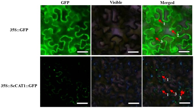
Figure 3. Subcellular localizations of ScCAT1 and empty vector in Nicotiana benthamiana leaves 48 h after infiltration.
The epidermal cells were used for taking images of green fluorescence, visible light and merged light. Read arrows 1, 2 and 3 indicated plasma membrane, nucleus and cytoplasm, respectively. Bar = 50 µm.

