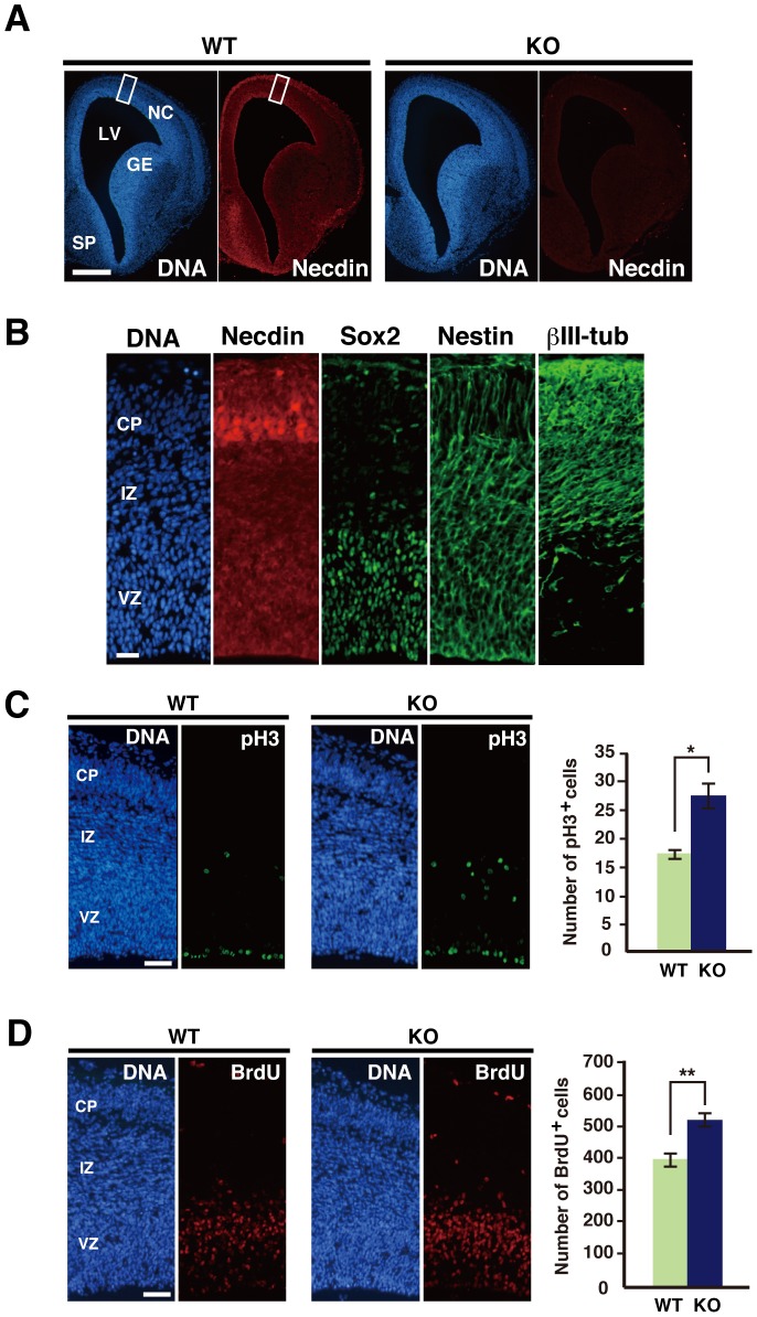
Figure 1. Necdin deficiency increases proliferative cell populations in the embryonic neocortex.
(A) Distribution of necdin in the forebrain. Cryosections were prepared from wild-type (WT) and necdin-null (KO) mice at E14.5 and immunostained for necdin. Abbreviations: NC, neocortex; GE, ganglionic eminence; LV, lateral ventricle; SP, septum. (B) Distribution of necdin, Sox2, nestin, and βIII-tubulin (βIII-tub) in the neocortex. The area shown is boxed in (A). Abbreviations: CP, cortical plate; IZ, intermediate zone; VZ, ventricular zone. (C) Phospho-histone H3 (pH3) immunohistochemistry. Cryosections were prepared from wild-type (WT) and necdin-null (KO) mice at E14.5, and immunostained for pH3. (D) S-phase cell population. Forebrain sections of E14.5 embryos were prepared 4 hrs after BrdU injection into pregnant female mice. BrdU+ cells in the neocortex were detected by immunohistochemistry. Chromosomal DNA (DNA) was stained with Hoechst 33342 in (A–D). pH3+ and BrdU+ cells within each 200-µm-wide radial column of the neocortex are counted (mean ± SEM, n = 3). *p<0.05, **p<0.01. Scale bars, 250 µm in (A), 100 µm in (B), 50 µm in (C, D).

