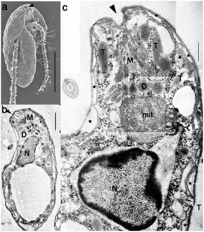
Figure 1. General morphology of Psammosa pacifica.
a. Surface structure of the ventral side of Psammosa pacifica, showing two flagella inserted subapical region of the ventral side. The opening of the gullet (arrowhead) is located at the cell apex. b. A longitudinal section of the cell showing a cluster of micronemes (M), dense vesicles (d) and the nucleus (N). c. The longitudinal section of the apical region shows a cluster of micronemes (M), dense vesicles (D) between the opening of the gullet (arrowhead) and a mitochondrion (mit). Single membrane bound trichocysts (T) are located near the cluster of micronemes. Alveoli vesicles (asterisk) are absent at the gullet and trichocysts. N: nucleus. Scales: 5 µm in a, 1 µm in b, 500 nm in c.

