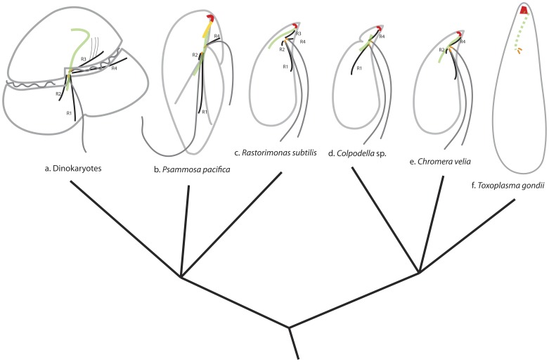
Figure 9. Comparison of the flagellar apparatus and the apical complex in myzozoans.
a. General dinokaryotes. b. Psammosa pacifica (this study). c. Rastorimonas subtilis [21]. d. Colpodella vorax [20]. e. Chromera velia [49], f. Toxoplasma gondii [16]. R1: root 1; R2: root 2; R3: root 3; R4: root 4. Green: bypassing microtubule strands (solid line) or SF-assemblin containing fibre (broken line); red: pseudoconoid or conoid; orange: basal bodies or centorioles.

