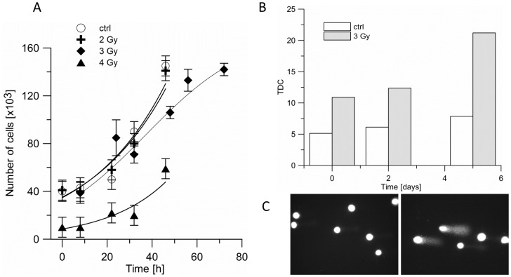
Figure 1. The effect of proton beam irradiation on BLM melanoma cells.
A. Proliferation of BLM cells after proton beam irradiation with doses 0 (open circle), 2 (cross), 3 (diamond) and 4 Gy (triangle). Cells were irradiated in suspension, and then plated in 96-well plates. B, C. DNA damage in untreated (white bar), and irradiated with 3 Gy of proton beam (stripped bar) BLM cells are presented in terms of the percentage of DNA that left the comet's head and was found in the comet’s tail after electrophoresis (TDC).

