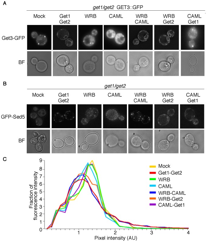
Figure 2. In combination, WRB and CAML rescue Get3 localization at the ER membrane and TA protein targeting.
(A) get1/get2 yeast cells carrying a genomically GFP-tagged version of Get3 were transformed with combinations of WRB, CAML, Get1 and Get2 encoding constructs. Subcellular Get3-GFP localization was analyzed by fluorescence microscopy. (B) get1/get2 yeast cells were transformed with a plasmid containing the coding sequence of GFP-tagged Sed5 and combinations of WRB, CAML, Get1 and Get2 encoding constructs. Subcellular GFP-Sed5 localization was analyzed by fluorescence microscopy. (C) Images taken in (B) were quantified to determine the distribution of fluorescence across bins of different pixel intensity for each strain. A minimum of 41 cells was analyzed per strain.

