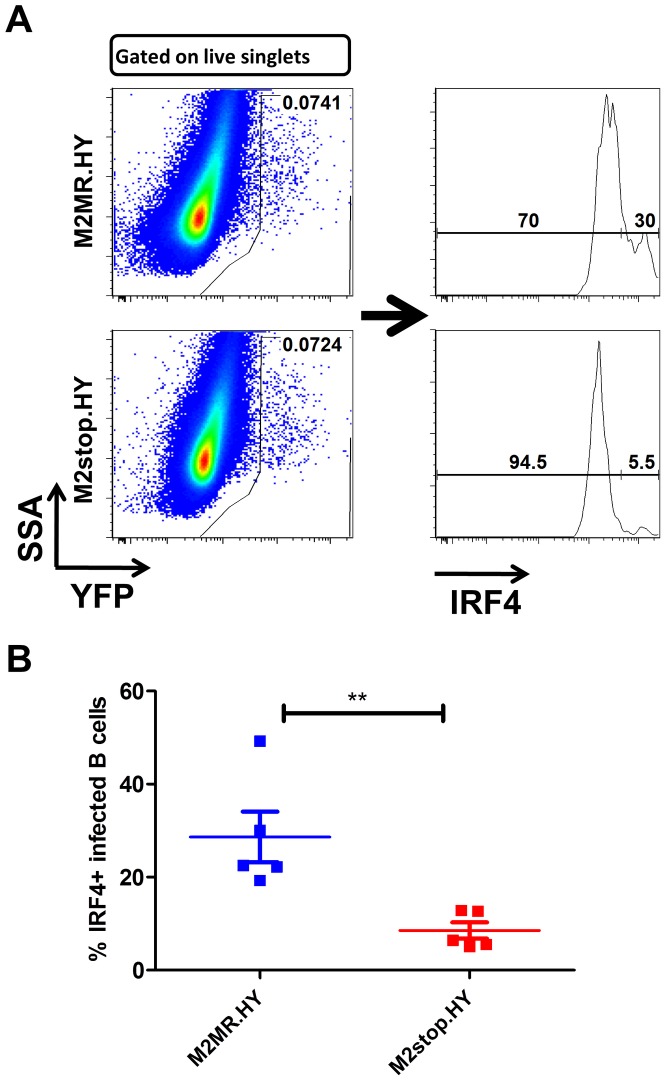
Figure 9. M2stop infected mice exhibit a decreased frequency of infected B cells expressing IRF4.
C57BL6/J mice were infected intraperitoneally with 1000 PFU of the indicated viruses and splenocytes were analyzed on day 14 post infection. (A) Representative flow plots illustrating the gating strategy. Splenocytes were gated to remove doublets following gating on the live cell population using a live/dead stain (Materials and Methods). (B) Frequency of live, singlet YFP+ cells that were IRF4 positive between M2stop.HY and M2MR.HY viruses. Data shown is from individual mice with five mice per virus group.

