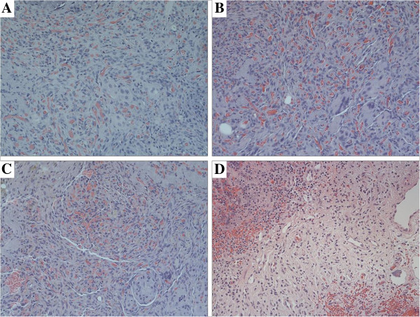
Figure 2.
Histopathology of xenograft AVMs. (HE × 200). 30 days (A), 60 days (B), 90 days (C) after transplantation, abundant of plump ECs with inconspicuous lumina were crowded together, in the center of the tissue. The densely of cells and vessels were almost maintained despite day of harvest. ECs in the grafts which are characterized by large nuclei and scant cytoplasm, lining inconspicuous vessels can be found has the same structural characteristics as the ECs in the pre-implanted human AVM nidus (D).

