
Fig. 1.
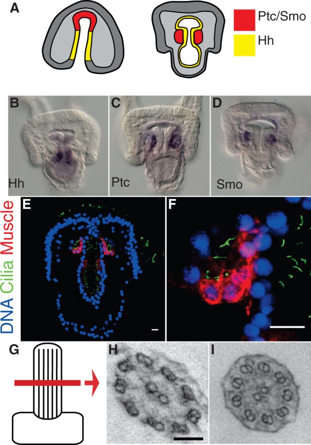
Hh-receiving cells exhibit motile cilia. (A) Model of Hh signaling in the sea urchin. Hh is secreted from the gut endoderm (yellow) and diffuses to the adjacent mesoderm where Ptc and Smo are expressed (red). (B–D) In situ mRNA hybridization of Hh (B), Ptc (C), and Smo (D). (E and F) Presumptive muscle cells display monocilia. (E) Hh-receiving cells are labeled by the muscle-specific MHC antibody (red), and cilia are labeled with an acetylated tubulin antibody (green). Scale bars = 10 μm. (F) Enlargement. (G–I) Transmission electron micrographs of cilia ultrastructure. (G) Cartoon representing the lateral region of the cilium scanned. (H) Cross-section of cilium from ectoderm showing 9 + 2 microtubule arrangement. Scale bar = 100 nm. (I) Cross-section of cilium on Hh-receiving cells of coelomic pouch also with a 9 + 2 arrangement.