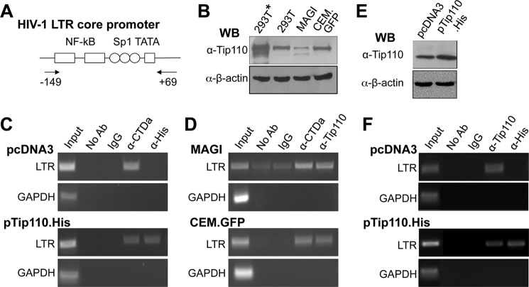
FIGURE 7.
Presence of Tip110 at the HIV-1 LTR core promoter. A, schematic of the LTR core promoter and the primer locations for PCR amplification. B, Western blot analysis for Tip110 expression in 293T cells transfected with pNL4-3 and pTip110.His (293T*) or pcDNA3 (293T), U373MAGI containing the LTR promoter-driven β-galactosidase gene (MAGI), and CEM-GFP cells containing the LTR promoter-driven GFP gene (CEM.GFP). C and D, 293T cells transfected with the pNL4-3 and pTip110.His (C, bottom) or pcDNA3 (C, top), U373MAGI (D, top), and CEM-GFP cells (D, bottom) were subjected to the ChIP assay. Following chromatin cross-linking, shearing, and immunoprecipitation with α-CTDa, α-His, or α-Tip110 antibody, reverse cross-linking was performed, and the DNA was then purified and analyzed by PCR with the primer set specific for the HIV-1 LTR core promoter region, as shown in A. Input DNA and immunoprecipitated DNA without any antibody (No Ab) or with an isotype-matched IgG were included as the ChIP controls. In addition, PCR using a primer set specific for a GAPDH coding region was performed and also included as the control. E and F, Jurkat cells were transfected with Tip110.His or pcDNA3 and then infected with NL4-3 (equivalent to 20,000 cpm of reverse transcriptase, which gave rise to about 80% infection in 3 days determined by intracellular p24 staining). Cells were harvested for Western blot analysis for Tip110 expression (E) or ChIP assay as described above (F).

