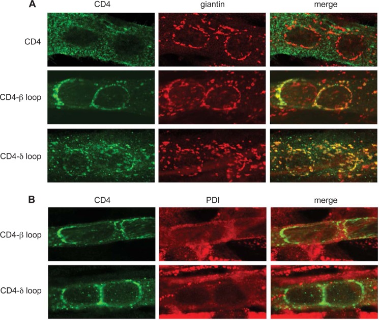
FIGURE 2.
CD4-β and δ cytoplasmic loops are retained mainly in the Golgi complex. A, C2 myotubes transfected with CD4 and CD4-β or δ loops were fixed, permeabilized, and double-stained with anti-CD4 antibody and an anti-giantin antibody to stain the Golgi complex. CD4 exhibited a diffuse intracellular and cell surface distribution, with little overlap with the giantin staining. In contrast, both CD4-β and δ loops exhibited extensive co-localization with the Golgi marker (see merged images). B, transfected myotubes were immunostained with anti-CD4 and an anti-protein disulfide isomerase (PDI) antibody to stain the endoplasmic reticulum. CD4-β or δ loops showed minimal co-localization with the ER marker. All confocal images are centered on a pair of nuclei within the multinucleate myotubes.

