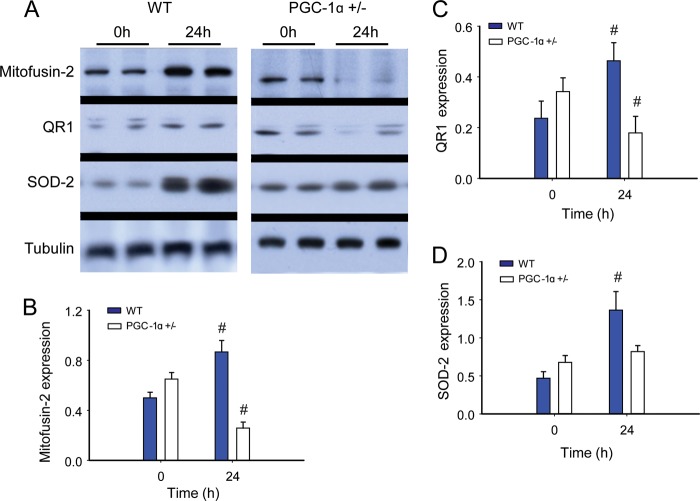
FIGURE 8.
Redox-regulated mitochondrial proteins during S. aureus sepsis in WT and PGC-1α+/− mice. A, Western blot analysis. Tubulin acts as a reference protein. B–D, protein densitometry for mitofusin-2 (B); QR1 (C); and SOD-2 (D) in WT and PGC-1α+/− mice at 0 h (control) and 24 h after clot implantation (n = 4). #, significant difference from 0 h.

