
FIGURE 6.
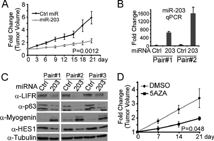
Re-expression of miR-203 inhibited tumor growth in nude mice. 1 × 10⁷ RD cells were first injected into both flanks of the nude mice to form solid tumors. A–C, control (Ctrl) miRNA or miR-203 was repeatedly injected into the tumors on each flank of the mice (n = 5) once every 3 days for a total of 21 days. A, diameter of tumors was measured, and the tumor volume (size) was calculated. The data are presented as mean ± S.D. B, total RNA was collected from two pairs of tumors and subjected to RT-qPCR analysis. Fold change was calculated as the ratio of the relative miR-203 level in the miR-203-treated tumors over that in the control miRNA-treated tumors. C, WCE were prepared from three pair of tumors, and an equal amount of WCE was subjected to SDS-PAGE and Western blot analysis. D, 5-aza-dC (5AZA) or DMSO was repeatedly injected into the tumors on each flank of the mice (n = 3) once every 3 days for a total of 21 days. The diameter of tumors was measured, and the tumor size was calculated. The fold change in A and D was calculated as the ratio of the tumor volume at different time points with miRNA or drug treatment over that before the treatment. The data were presented as mean ± S.D.