FIGURE 5.
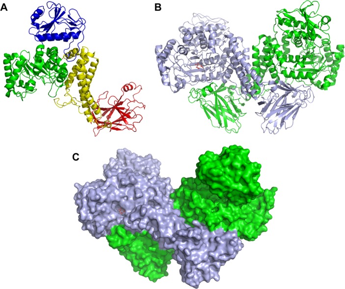
Structure of BoAgu115A. Panel A is a schematic of a protomer of BoAgu115A showing the four distinct domains of the enzyme: the N-terminal β-strand/α-helix domain (blue), the core TIM barrel catalytic domain (green), the five-helical bundle domain (yellow), and the C-terminal β-sandwich domain (red). Panel B depicts the enzyme in its dimeric butterfly-like form with protomers 1 and 2 colored green and light blue, respectively. Panel C shows a surface representation of the GH115 dimer. The relative orientation of the dimer and color of the protomers is as indicated in B. Both panels B and C show the GlcA bound form (sugar shown in stick format, carbons colored salmon pink).

