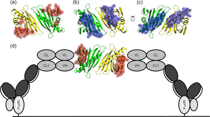
FIGURE 7.
Accessibility of IgE epitopes in the tetrasulfide dimer. Residues corresponding to IgE epitopes are highlighted as sticks and surface. a, b, and d, three of four reported epitopes allow cross-linking of univalent IgE antibodies via dimeric Bet v 1 (shown as red surface) (38, 40). c, by contrast, a fourth epitope at the C-terminal α-helix of Bet v 1 (blue surface) is partly buried by the dimer interface, restricting simultaneous binding of two IgE (39). d, a schematic model for IgE cross-linking facilitated by dimeric Bet v 1 on the surface of an effector cell, mediated by FcϵRI, based on Ref. 7.

