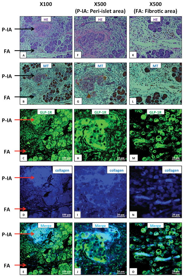
Figure 4. Glucagon-like peptide-1 receptor (GLP-1R) and type I collagen expression in pancreas of Wistar/Bonn-Kobori (WBN/Kob) rats (a chronic pancreatitis model).

Shown are representative sections showing peri-islet area (P-IA) and a fibrotic area (FA) at the indicated magnifications (100x, 500x). Expression of GLP-1R and type I collagen in the pancreas of 15-week-old WBN/Kob rats was examined by immunofluorescence staining. Panel (A, F, K) shows Hematoxylin-Eosin (HE) staining, Panel (B, G, L) Masson Trichrome (MT) staining for collagen fibrils, Panel (C, H, M) GLP-1R staining (green), Panel (D, I, N) type I collagen staining (blue) and Panel (E, J, O) a merged image of type I collagen and GLP-1R. These pictures are from sequential sections showing the same area. This figure demonstrates in this chronic pancreatitis model increased collagen deposition (MT staining) and increased GLP-1R expression in areas where increased collagen is present (merged picture). These pictures are representative images from four experiments.
