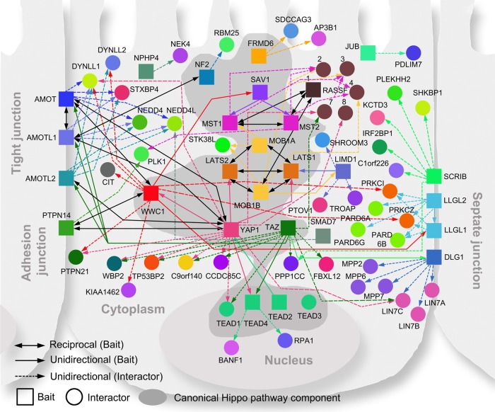
Fig. 3.
Protein–protein interaction network of the Hippo pathway. Canonical Hippo pathway components are highlighted with a dark gray background. The unidirectional (solid colored and single arrows) and reciprocal (solid black and double arrows) interactions between baits (squares) are shown. Each bait protein is rendered in a unique color and linked to its major HCIPs (circles) by a dashed line with a single arrow in the corresponding color.

