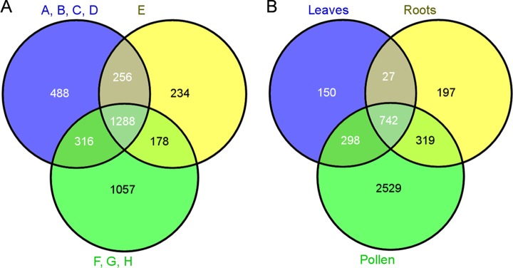
Fig. 3.

Venn diagrams. A, number of proteins identified throughout development. A–D, microsporocytes, meiotic cells, tetrads, and microspores, respectively; E, polarized microspores; F–H, binuclear pollen, desiccated pollen, and pollen tubes, respectively. B, number of identified proteins in all pollen stages, leaves, and roots.
