Fig. 1.
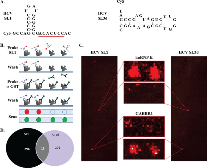
Multiple host proteins bind to SL1 in the HCV genome. A, Sequences and putative structures of SL1 and SL3d used to identify interacting proteins in human protein microarrays. SL1 (nt 1–30 of the HCV genome; left panel) and SL3d (nt 251–280 of the HCV genome; right panel) were both synthesized to contain a Cy5 dye at their 5′ termini. The seed sequence for miR-122 binding in SL1 is underlined. B, Schematic of the screening process for the protein microarrays. The RNAs are shown as hairpins with a red circle to denote Cy5. The DyLight™549-conjugated anti-GST antibody used to assess the amount of each protein spot is shown as a Y with a green circle. C, Two examples of hits from the human protein arrays probed respectively with the HCV SL1 (left panel) and HCV SL3d (right panel). The central panels show the spots corresponding to hnRNP K and GABBR1 and how they specifically bind to HCV SL1 and SL3d, respectively. D, Summary of the hits from the screens. In total 313 and 224 proteins were found to bind HCV SL1 and SL3d, respectively. Note that 19 proteins bind to both RNAs.

