Fig. 5.
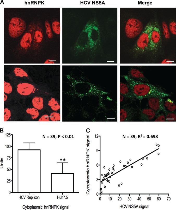
HCV replication increases hnRNP K abundance in the cytoplasm. A, Immunofluorescence analysis of representative Huh7.5 cells with or without HCV NS5A-GFP replicon (20). Four representative cells that contained the HCV replicon (two in each of the upper and lower panels) are shown. The endogenous hnRNP K was visualized with anti-hnRNP K antibody and Alexa Fluor 594-conjugated secondary antibody. The NS5A protein, in the NS5A-GFP replicon, was detected via GFP fluorescence. hnRNP K was observed in all nuclei; it was also found as the hazy red color in the cytoplasm of the four representative cells. Note that NS5A subcellular distribution (middle and right-most panels) correlates with the hnRNP K fluorescence. B, Quantification of the amount of cytoplasmic signal (red) for hnRNP K in 39 independently selected Huh7.5 cells with or without HCV NS5A-GFP replicon. Quantification of the cytoplasmic fluorescence was performed with the Image J program. Statistical analysis was performed with the Student t test. C, A correlation between the level of NS5A-GFP expression and the abundance of hnRNP K presented in the cytoplasm.

