
Figure 1.
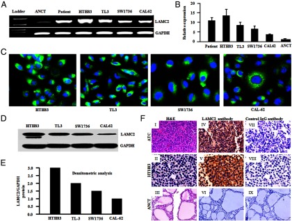
LAMC2 mRNA and protein expression in ATC cell lines and ATC tissue specimens. A, RT-PCR analysis demonstrated strong LAMC2 mRNA expression in human ATC cells (HTH83, TL3, SW1736, CAL62, and ATC351) and ATC tissue samples, whereas low LAMC2 mRNA expression was detected in the normal thyroid (ANCT) tissue sample. Glyceraldehyde-3-phosphate dehydrogenase (GAPDH) was used as an internal control. B, Real-time PCR analysis showed the relative expression of LAMC2 in ATC cell lines and ANCT. Data represent the mean ± SD of three independent experiments. C, Indirect immunofluorescence assay detected endogenous LAMC2 protein expression in fixed/permeabilized ATC cell lines; 4′,6′-diamino-2-phenylindole (DAPI) stains nuclei. D, Western blot analysis of ATC cell line demonstrated LAMC2 antibody specifically recognizes a band of 150 kDa corresponding to LAMC2 protein. E, Densitometric analysis of Western blot showed LAMC2 protein expression in ATC cell lines. F, Immunohistochemical analysis of ATC specimen and HTH83 cell line for the detection of LAMC2 protein. I, ATC sample. II, HTH83. III, ANCT: sections stained with hematoxylin and eosin (H&E). Cytoplasmic localization of the LAMC2 was observed in ATC specimen (IV) and HTH83 cells with anti-LAMC2 antibody (V), whereas ANCT tissues showed no reactivity (VI). Negligible staining was observed in the serial sections of the ATC sample (VII), HTH83 (VIII), and ANCT sample probed with control IgG (IX) (original magnification, ×200).