
Figure 5.
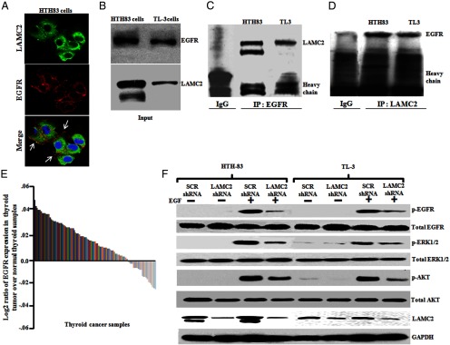
LAMC2 can bind to EGFR, and LAMC2 shRNA affected phosphorylation of EGFR and its downstream signaling in human ATC cells (HTH83 and TL3). A, HTH83 cells were fixed, permeablized, and immunostained, and images were captured using a confocal microscope. Alexa 488 (green fluorescence) and Alexa 680 (red fluorescence) were used for LAMC2 and EGFR staining, respectively. The images were merged for the colocalization of LAMC2 and EGFR. Arrows depict the colocalization. 4′,6′-Diamino-2-phenylindole was used to stain the nuclei of the cells. Images are representative of at least four independent experiments yielding similar results. Original magnification, ×600; objective, ×60. B, Western blot (WB) analysis showed endogenous levels of LAMC2 and EGFR protein in ATC cells (HTH83 and TL3). C, Cell lysates were subjected to immunoprecipitation using EGFR monoclonal antibody, and immunoprecipitates were Western blotted with LAMC2 monoclonal antibody. D, LAMC2 antibody was used for immunoprecipitation, and the immunoprecipitates were immunoblotted with EGFR monoclonal antibody. E, EGFR expression in thyroid cancer samples using microarray database (GSE27155). F, ATC cells stably expressing either LAMC2 shRNA or SCR shRNA were serum starved for 24 hours and then treated with EGF (50 ng/mL) for 20 minutes and phosphorylation of EGFR, ERK1/2, and AKT was analyzed by Western blot using phosphorylated (p)-EGFR, p-ERK1/2, and p-AKT antibodies. Glyceraldehyde-3-phosphate dehydrogenase (GAPDH) antibody was used for equal loading control.