
Figure 6.
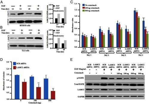
Induction of LAMC2 expression by EGF. Silencing LAMC2 enhanced the antiproliferative activity of ATC cells in combination with cetuximab. A and B, HTH83 and TL3 cells (106) cells were seeded into 10-cm culture dishes and serum starved for 24 hours followed by the addition of fresh. After serum starvation, cells were incubated either with fresh medium containing either EGF (50 ng/mL) or diluent control. The cells were harvested at the indicated time points for Western blotting. Densitometry analysis showed fold change in LAMC2 expression upon EGF treatment. C, HTH83 cells stably expressing either LAMC2 shRNA or SCR shRNA were seeded into 96-well plates and cetuximab (100 or 500 ng/mL) was added. The rate of proliferation was measured every 24 hours by a 3-(4,5-dimethylthiazol-2-yl)-2,5-diphenyltetrazolium bromide assay. D, HTH83 cells (2 × 10³) stably expressing either LAMC2 shRNA or SCR shRNA were used for a clonogenic growth assay in the presence of cetuximab (100 or 500 ng/mL). For panels C and D, results represent the mean ± SD of three independent experiments. *, P ≤ .01; **, P ≤ .001 (Student's t test). E, Western blot (WB) analysis. HTH83 LAMC2 shRNA cells and HTH83 SCR shRNA cells were treated with cetuximab (100 or 500 ng/mL) for 24 hours and then serum starved for 24 hours followed by the addition of EGF (50 ng/mL) for 20 minutes, and phosphorylation (p) of EGFR was analyzed by Western blot using antibodies against p-EGFR, EGFR, LAMC2, and glyceraldehyde-3-phosphate dehydrogenase (GAPDH; loading control).