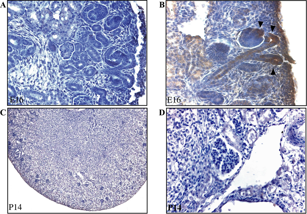
Figure 5. The pattern of immunohistochemical staining for β-galactosidase conforms to the pattern of TCF/β-catenin-lacZ reporter activity.
Wildtype embryos bearing the TCF/β-catenin-lacZ reporter were subjected to immunohistochemistry for β-galactosidase without (A) and with (B) the addition of the primary antibody at embryonic day E16. Arrowheads show location of β-galactosidase in UB and comma-shaped body. At 2 weeks of age, no immunostaining for β-galactosidase was evident in the normal or cystic compartments of Pkd2/WS25 kidneys. Representative images at 10X magnification (A–C) and 40X magnification (B–D) are shown.

