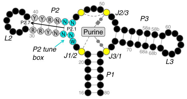
Fig. 1.

General schematic of the guanine/adenine riboswitch aptamer domain. Nucleotides are shown as black circles, excluding the P2 tune box (cyan), P2 core (gray), and purine-binding pocket (yellow). Bases are numbered according to a scheme in Stoddard et al. The numbering scheme for P2 is labeled (P2.1–P2.7). A purine is represented in the binding pocket with conserved bonds shown. Base pairs with the ligand are drawn using the Leontis–Westhof notation while single hydrogen bonds are shown as gray broken lines. The figure was generated using Varna.12
