
Fig. 1.
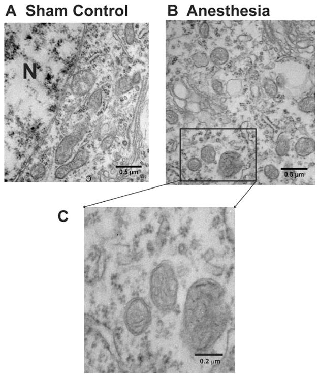
Anesthesia causes acute ultrastructural changes in mitochondria of pyramidal subicular neurons of 8-day-old rats. (A) Mitochondria in the cytoplasm of subicular pyramidal neurons from sham control animals resemble long tubules with intact inner and outer membranes and numerous cristae tightly packed inside healthy looking matrix. (B) Mitochondria in the cytoplasm of subicular pyramidal neurons from anesthesia- treated animals are numerous. The mitochondria are round, small, and display globular morphology 24 h postanesthesia exposure (on P8). Their matrix is pale and shows the signs of swelling. Although the inner and outer membranes appear somewhat intact, the cristae seem distorted and difficult to discern (C). N = nucleus.