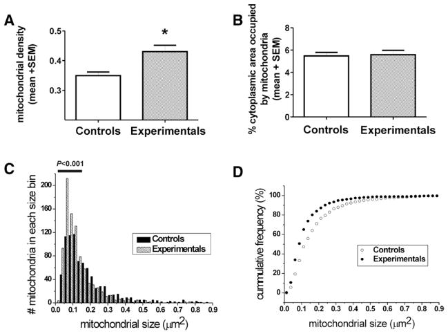
Fig. 2.
Anesthesia induces excessive fission of mitochondria in the soma of pyramidal subicular neurons of 8-day-old rats. (A) Mitochondrial density was assessed by counting the number of mitochondrial profiles per unit area (μm2) of cytoplasmic soma in each pyramidal neuron. There are approximately 30% more mitochondrial profiles in anesthesia-treated neurons compared with controls (*P = 0.0179). (B) The summation of mitochondrial areas, presented as a percent of the cytoplasmic area of pyramidal neurons, reveals that mitochondria in the control and experimental neurons occupy approximately the same area of the cytoplasmic soma (P = 0.8067). (C) Frequency distribution analysis by grouping of mitochondrial area indicates that there are significantly more mitochondria smaller than 0.16 μm2 (indicated with horizontal bar) in experimental animals compared with controls (P < 0.001). (D) Cumulative frequency analysis (in percentage), designed to take into account the differences in overall mitochondrial number in control vs. experimental neurons, indicates a leftward shift toward smaller mitochondria after anesthesia treatment, with over 50% of mitochondria in the category of lesser than 0.1 μm2. In addition, mitochondria smaller than 0.012 μm2 were detected in the anesthesia-treated pyramidal neurons, whereas none that small could be detected in the control neurons (n = 4 control and four experimental pups, five neurons from each pup).

