
Fig. 1.
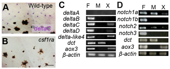
Expression analysis of Notch receptors and their ligands. (A,B) In situ hybridization for deltaC in larval fish. Presumptive xanthophores in the inter stripe are stained in wild type (A) but these cells and deltaC staining is absent in the csf1ra mutant (B). (C,D) Expression by RT-PCR of major Notch ligands (C) and Notch receptors (D). F, fins; M, melanophores; X, xanthophores. dct and aox3 are markers for melanophores and xanthophores, respectively. Scale bar: 50 μm.