Fig. 1.
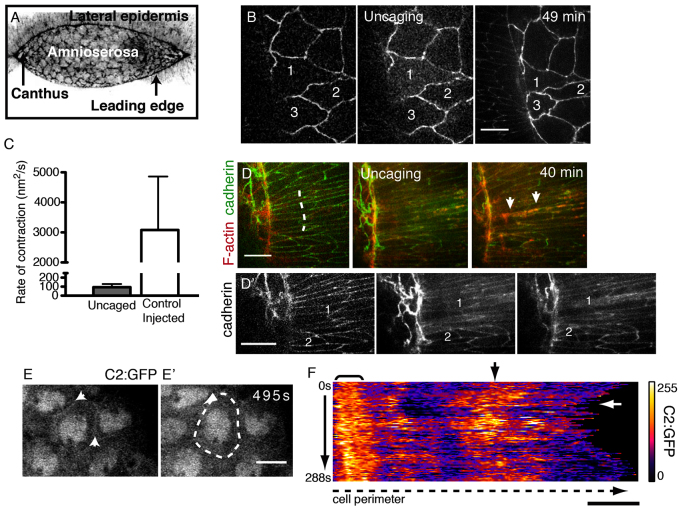
Calcium triggers rapid cell contractions. (A) Confocal micrograph (reverse contrast) of closure in a wild-type Drosophila embryo. (B) Uncaged Ca2+ induces constriction in the AS of an embryo expressing E-cadherin-GFP and GCaMP3. Targeted cells are numbered for reference. (C) Rate of contraction in uncaged versus control AS cells. Error bars indicate s.d. (D) Uncaged Ca2+ induces apical F-actin protrusions (red, arrows) in targeted LE (dashed line). (D′) Cadherin signal alone. (E,E′) An AS cell expressing C2:GFP; arrows indicate cell boundaries. (F) Kymograph of GFP signal along the traced boundary (dashed line in E′) over time (3 seconds/frame). Bracket indicates persistent C2:GFP signal; black arrow indicates a region with dynamic C2:GFP signal; white arrow indicates constriction (decreased cell perimeter). Scale bars: 10 μm.

