Figure 4.
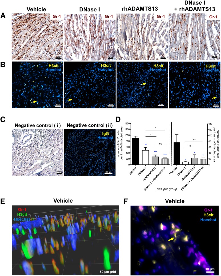
Treatment with DNase I and rhADAMTS13 significantly reduces leukocyte infiltration and H3cit accumulation in the infarcted area. (A) Immunohistochemical staining revealed abundant presence of Gr-1–positive cells (brown) infiltrating the ischemic myocardium in vehicle-treated mice and significantly less in heart sections of mice treated with DNase I monotherapy, rhADAMTS13 monotherapy, or DNase I in combination with rhADAMTS13. (B) Immunofluorescence analysis showed H3cit-positive cells in the infarcted area (yellow arrows). Numerous H3cit-positive cells were found in the vehicle-treated group, whereas only a few H3cit-expressing cells were found after treatment with DNase I, rhADAMTS13, or a combination of DNase I and rhADAMTS13. (C) Incubation with only secondary antibody served as a negative control (i). Negative control was performed using specific IgG instead of primary antibody (ii). (D) Numbers of cells positive for Gr-1 (left) and H3cit (right) were counted (4 mice per group) and presented as positive cells per 1 mm2 of infarcted area, respectively (*P < .05, **P < .01, ***P < .001; blue asterisks represent comparison with vehicle, and black between treated groups). (E) Representative deconvolved 3D image of double staining for Gr-1 (red) and H3cit (green) of the infarcted myocardium in a vehicle-treated mouse. Most H3cit-positive cells are associated with Gr-1 antigen. (F) Double immunofluorescence analysis for Gr-1 (purple) and H3cit (yellow). Yellow arrow indicates NET-like H3cit-positive structure. Scale bars: 50 μm (A-B, Ci), 200 μm (Cii), 50-μm grid (E), 10 μm (F).

