. 2013 Nov;21(6):481–486. doi: 10.4062/biomolther.2013.076

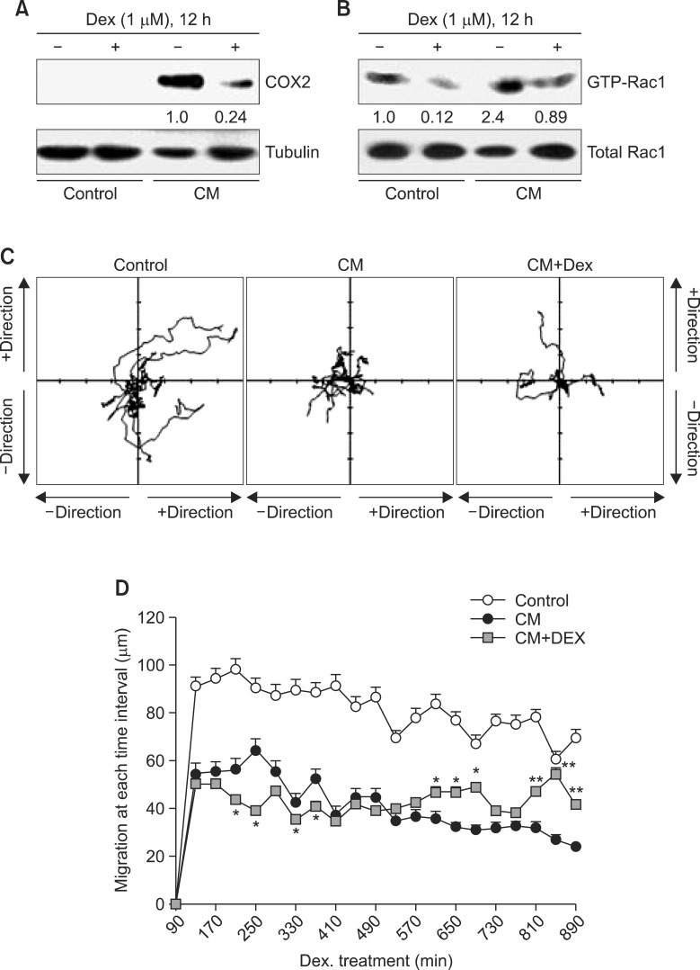
Fig. 4. CM-induced decrease in macrophage motility was reversed by the treatment with dexamethasone. (A) and (B) IC-21 cells were treated with CM in the presence or absence of dexamethasone (Dex). COX2 proteins were detected by immunoblot analysis (A). Rac1 activity was measured by GST-Pulldown assay as described in materials and methods (B). Band intensity to control protein was normalized with NIH image analysis software (version 1.62). Fold increase to control group was represented under each band (A and B). Macrophage motility was measured and the changes in image of each macrophage was analyzed with tracking program, ImageJ plugin MTrackJ (Version 1.5.0) by the method described in materials and methods. Movement track was presented in coordinates for each cell (C). Moved distance of macrophages was calculated at every 40 min interval for 15 h. Data were the representative of four experiments. Data in the bar graph represent the means ± SED. *p<0.05; **p<0.01, significantly different from CM-treated group (D).

Fig. 4.