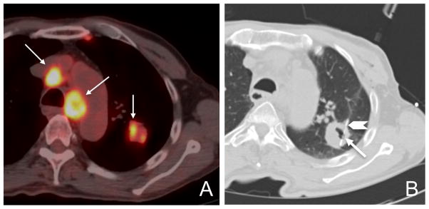
Figure 1.

Case of sampling error resulting in insufficient tissue for biomarker analysis. Axial fused PET/CT image demonstrates FDG activity (arrows) within the mediastinal nodes and the inferior, medial border of the left upper lobe lung mass (A). Axial CT image during lung biopsy to acquire tumor tissue for enrollment into BATTLE demonstrates a coaxial technique with the tip of the 19-gauge guide needle located at the periphery of the lesion (arrowhead) and the 20-gauge core biopsy needle located in the lateral edge of the cavitary mass (arrow), an area which does not correspond to the highest area of FDG uptake on the pre-biopsy diagnostic PET/CT (B).
