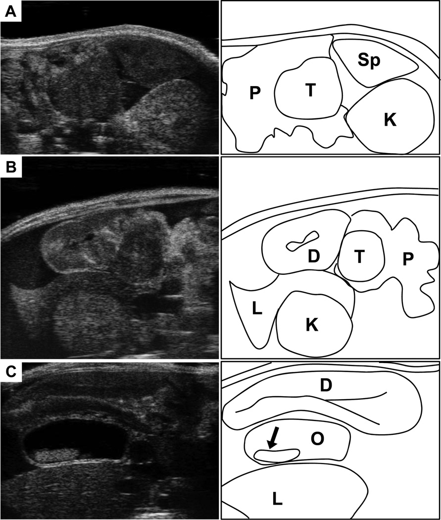
Figure 2. Landmarks for location the mouse pancreas.
(A) Ultrasound image (left) of the tail of the pancreas (P), with a solid pancreatic tumor (T). Note location of the pancreas relative to the spleen (Sp) and left kidney (K). (B) The head of the pancreas (P) with a solid pancreatic tumor. This region can be more complicated to discern than the tail of pancreas. Landmarks include the proximal duodenum (D), which can be traced extending from the pylorus of the stomach. Also note the right kidney (K) and the right lobe of the liver (L). (C) Another common feature observed with head of pancreas tumors is the development of common bile duct obstructions (O). Here the bile duct is dramatically enlarged, and stones have developed (arrow). Nearby landmarks seen here include the duodenum (D) and the right lobe of the liver (L).

