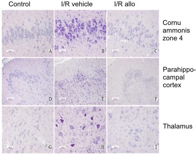
Figure 2.
Histopathological images; acid fuchsin thionin staining. Representative images of the histopathological appearance of fetal sheep brains at 0.8 of gestation in controls (A, D, and G), the ischemia–reperfusion (I/R) vehicle group (B, E, and H), and the I/R allopurinol group (C, F, and I). Panels A to C represent zone 4 of the cornu ammonis; panels D to F are images of the parahippocampal cortex; and panels G, H, and I represent the thalamus. An acid fuchsin and thionin staining was used to detect neuronal loss (acidophilic positive neurons).

