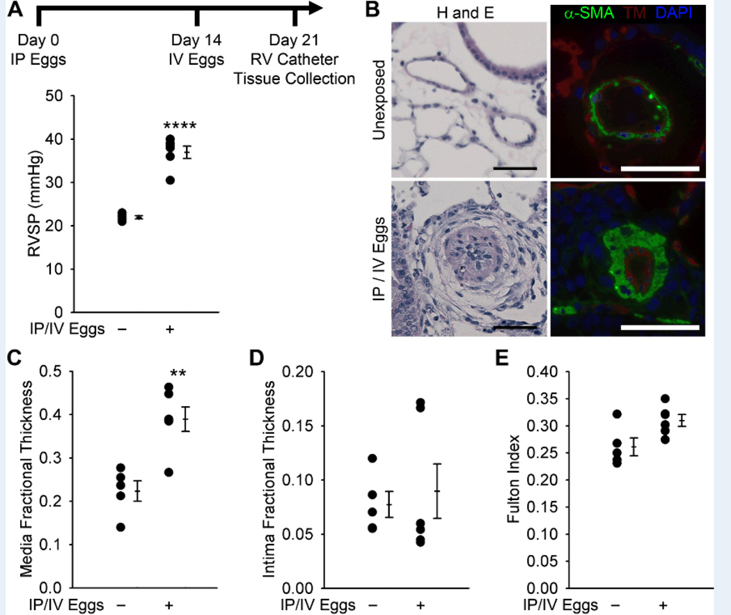
Figure 1.
Mice exposed to S. mansoni eggs develop PH and vascular remodeling. (A) Mice IP sensitized to S. mansoni eggs followed by IV augmentation have an increase in RVSP (mean ± SE; n = 5–6 mice per group; rank-sum test ****P < 0.001; this experiment was repeated 3 times with similar results). (B) Representative H&E and immunofluorescence staining for α-smooth muscle actin (α-SMA) and thrombomodulin of unexposed and IP/IV egg exposed mouse lungs (Scale bars: 50µm). (C and D) Quantitative fractional thickness of the pulmonary vascular media and intima in unexposed and IP/IV egg-exposed mice (mean ± SE; n = 5–6 mice per group; rank-sum test **P < 0.01; P=0.66 for intima thickness). (E) Fulton index (RV/(LV+S)) of unexposed and IP/IV egg exposed mice (mean ± SE; n = 5–6 mice per group; rank-sum test P = 0.052).

