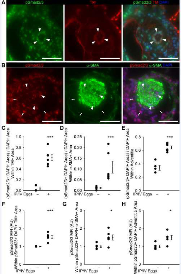
Figure 5.
Quantification of phospho-Smad2/3 and co-localization with vascular compartments in unexposed and mice exposed to IP/IV S. mansoni eggs. (A) Representative images showing phospho-Smad2/3 and thrombomodulin (TM) co-localization in the intima of an egg-exposed mouse (arrowheads mark double-positive cells; Scale bars: 50 µm). (B) Representative images showing phospho-Smad2/3 and α-smooth muscle actin (α-SMA) co-localization in the media, and identifying the adventitia, of an IP/IV S. mansoni egg-exposed mouse (arrowheads mark double-positive cells in the media; arrows mark phospho-Smad2/3+ cells in the adventitia; Scale bars: 50 µm). (C–E) Quantification of phospho-Smad2/3+ thrombomodulin+ area in the intima, media and adventitia (mean ± SE; n = 5–6 mice per group; rank-sum test ***P<0.005). (F–H) Mean fluorescent intensity of phospho-Smad2/3+ pixels in the initima, media and adventitia (arbitrary units; normalized to average of uninfected = 1; mean ± SE; n = 5–6 mice per group; rank-sum test *P<0.05, ***P<0.005).

