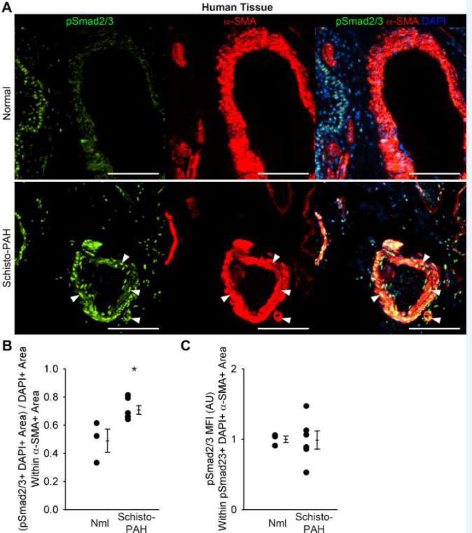
Figure 6.
Human schistosomiasis-PAH tissue analysis by phospho-Smad2/3 quantification and co-localization with the media vascular compartment, as compared to control tissue from failed lung donors. (A) Representative images showing phospho-Smad2/3 and α-smooth muscle actin (α-SMA) co-localization in the media of normal control and Sc-PAH cases (arrowheads mark double-positive cells; Scale bars: 100 µm). (B) Quantification of phospho-Smad2/3 α-SMA double-positive area in the media (mean ± SE; n = 3–6 cases per group; rank-sum test *P<0.05). (C) Mean fluorescent intensity of phospho-Smad2/3+ pixels in the media (arbitrary units; normalized to average of control = 1; mean ± SE; n = 3–6 cases per group; rank-sum test P=1.0).

