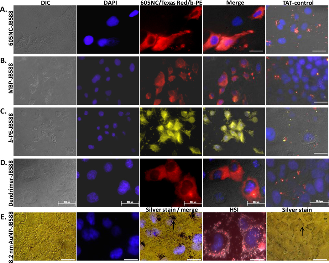
Figure 4. JB577-mediated cytosolic delivery of disparate protein and nanoparticle materials.
JB588 peptide EDC-conjugated to (A) eBioscience 605NC (5 nM),33 (B) Texas Red-labeled maltose binding protein (200 nM), (C) B-phycoerythrin (5 nM),54 (D) Tetramethylrhodamine isothiocyanate-(TRITC) labeled G5-PAMAM dendrimer (50 nM) and (E) 8.2 nm gold NPs (100 nM, silver stained) and delivered to COS-1 cells as described. All show cytosolic dispersal. Gold NPs localized in what appear to be perinuclear spaces as indicated by the arrow (under Silver stain / merge). See right column of images where a control CPP- (JB586-TAT, Arg7) conjugate of each species consistently showed NP sequestration within endosomes or alternatively lack of uptake for the gold NPs (arrow under Silver stain indicates AuNPs located at the cell periphery). HIS is hyperspectral imaging which combines simultaneous dark field and fluorescence imaging.36 In all cases, NP materials were incubated with cells for 1–2 h, washed and cells cultured for 48 h prior to fixation. Magnification: 60x (b, c); 100x (a, d, e). Size bar = 50 µm except for HSI which is 100 µm

