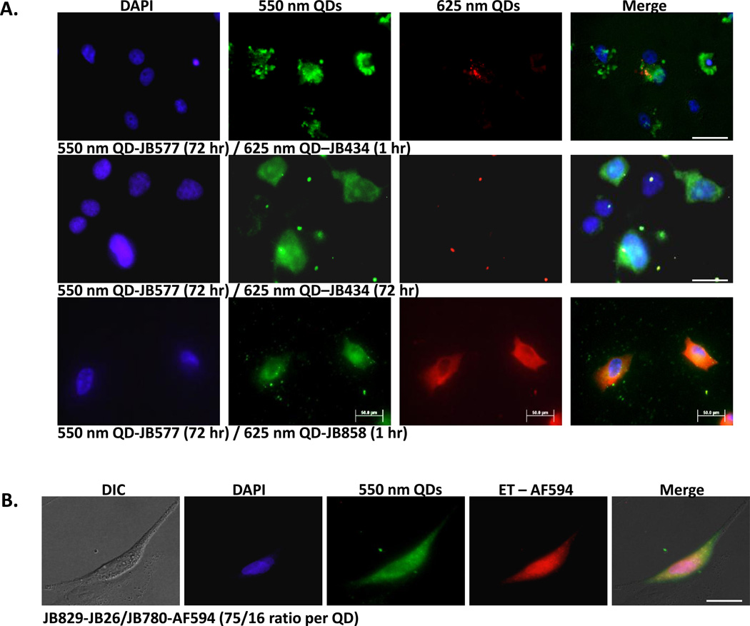
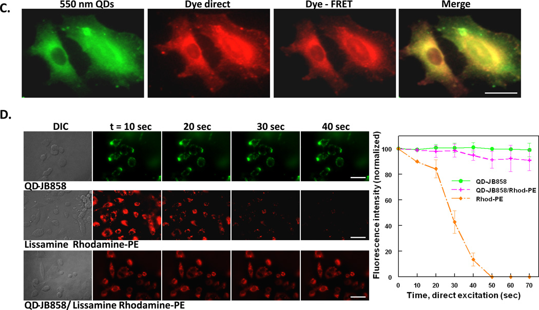
Figure 7. Combinatorial QD-peptide cellular labeling and improved membrane visualization by sensitization.
(A) Combinatorial labeling. COS-1 cells labeled with 100 nM 550 nm QDs (75-JB577 peptides/QD; cytosol) and 2 nM 625 nm QDs (20 JB434 peptides/QD; endosomes) by either sequential (top row) or simultaneous incubation (middle row) of the QD-peptide conjugates. Sequential delivery consisted of initial 2 h incubation with QD-JB577 complexes, 72 h cell culture followed by 1 h incubation with QD-JB434 assemblies. For simultaneous incubation, both QD-peptide complexes were incubated on cells for 2 h, removed and cells cultured for 72 h followed by fixation and DAPI-staining. (Bottom row) Sequential labeling of cytosol and plasma membrane of A549 cells with 150 nM 550 nm QDs (75 JB577 peptides/QD, cytosol) and 10 nM 625 nm QDs (75 JB858 peptides/QD, membrane). QD-JB577 assemblies were incubated for 3 h, removed and cells cultured 72 h followed by 1 h incubation with QD-JB858 conjugates prior to fixation. Lower 625 nm QD concentrations are used due to the 3-fold higher Q.Y. (B) Cytosolic QD-peptide cargo delivery and stability. A549 cells were labeled with 550 nm QDs (100 nM) assembled with peptides JB829-26 (75/QD) and AlexaFluor (AF) 594-labeled JB780 (16/QD). QD-peptides were incubated on cells for 3 h followed by 72 h culture period prior to fixation. AF594 is FRET sensitized from direct excitation of the 550 nm QD donor (Förster distance/R0 ~3.7 nm). (C) Plasma membrane of PC12-Adh cells were labeled sequentially with 125 nM 550 nm QD-JB858 conjugates (1 h incubation) followed by 20 min incubation with 10 µM Lissamine Rhodamine B phosphoethanolamine (Rhod-PE). The dye-FRET panel shows sensitization of the dye by the QD donor (R0 ~5.2 nm). (D) Comparison of the photostability of membrane resident QDs, Lissamine Rhodamine B phosphoethanolamine, and the QD-Rhodamine bi-labeled cells. Sensitization circumvents direct dye photobleaching. Full experimental details are given in the SI. Size bar = 50 µm


