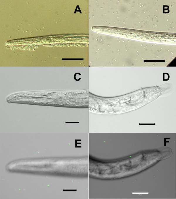
Figure 3.
Observation of Serratia sp. LCN-16 in association with Bursaphelenchus xylophilus after 1 h and 24 h contact. (A, B) Differential interference contrast (DIC) microscope images of B. xylophilus, treated by 1 h contact of bacteria before (A) and after (B) washing with sterile DW. (C-F) DIC and fluorescence-merged images of B. xylophilus, treated by 24 h contact of bacteria and washed with sterile DW. The images of the head (C) and tail (D) region were captured in a single focal plane . Serial-section images were acquired and stacked, showing surfaces of the head (E) and tail (F) region. Scale bars, (A), (B), 30 μm; (C)-(F), 20 μm.

