Figure 1.
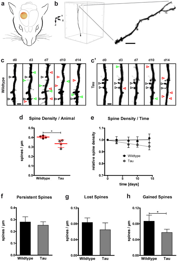
Decreased dendritic spine density and impaired spine kinetics in P301S Tau mice. a Location of the chronic cranial glass window, as implanted for long-term in vivo imaging of YFP-expressing neurons. b Example of an isolated, volume rendered layer V neuron in a 425 × 425 × 550 μm3 xyz-stack of the somatosensory cortex. A section of an apical tufted dendrite, stretching in parallel to the brain surface, is shown in higher magnification (maximum intensity projection; scale bar: 5 μm). c-c’ Dendritic elements in 4-month-old wildtype (c) and P301S Tau mice (c’) were analyzed by high-resolution two-photon in vivo imaging over a time period of two weeks (d: day). Repetitive imaging allows discrimination of stable, gained, and lost spines (black, green, and red arrowheads respectively; maximum intensity projections; scale bars: 2 μm). d Spine density was reduced in P301S Tau mice at the first in vivo imaging session. Presented are means per animal of 8 mice; 61–82 dendrites per group; 14–22 dendrites per mouse; means ± SD per group in red. e During the following 14 days, the spine density stayed stable in wildtype mice but further declined in P301S Tau mice. Presented are means ± SEM of 28–41 dendrites in 3–4 mice per group, 8–12 dendrites per mouse; normalized to the first imaging day. Line, linear fit. p <0.01 (F test of means). f-h In P301S Tau mice, the densities of persistent (f) and lost spines (g) was not significantly impaired, while the density of gained spines was decreased (h). Presented are means ± SD of 3–4 mice per group; 28–41 dendrites; 8–12 dendrites per mouse. b, c, e, f &g: Unpaired t test; * p < 0.05.

