
Figure 10.
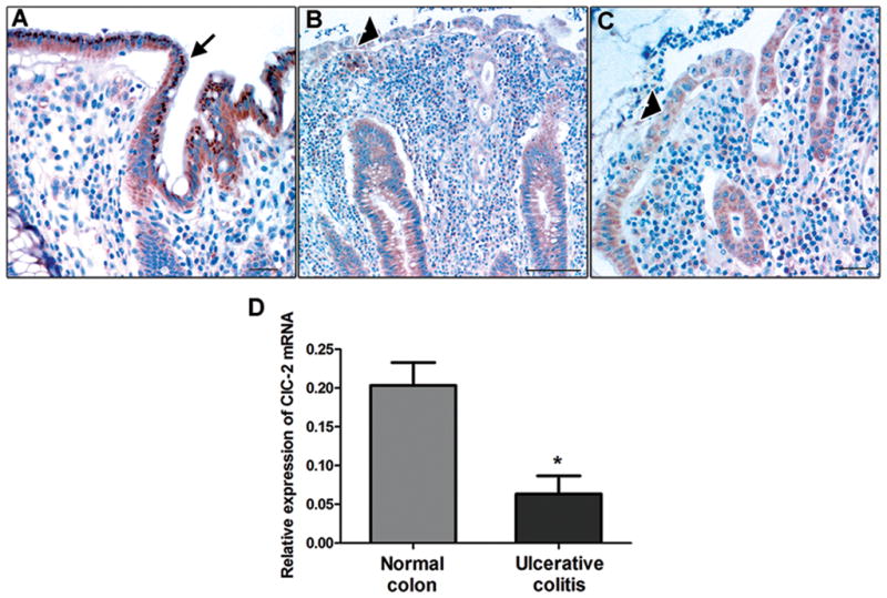
Immunohistochemistry and qPCR for ClC-2 in ulcerative colitis biopsies. A: In unaffected colonic tissue, ample ClC-2 staining was present (brown color, arrow). However, in UC patients, expression of ClC-2 in the colonic surface epithelium was found to be markedly reduced (B and C, arrowhead). Bar = 25μM. The images are representative of 6 normal and 6 UC biopsies. D: In the qPCR analysis, mRNA expression of ClC-2 was found to be significantly reduced in UC colonic biopsies compared to control patients. The ClC-2 mRNA expression is normalized to mRNA expression of GAPDH. (n = 3 in each group, *p < 0.05).