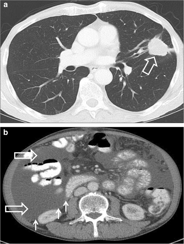
Figure 2.

Contrast-enhanced computed tomography scan of the chest and abdomen showing disease progression in June 2011. (a) A 3.3 × 2.8 cm mass (big arrow) in the left lung and (b) ascites in all 4 quadrants of the abdomen (big arrow) and peritoneal nodules (small arrows).
