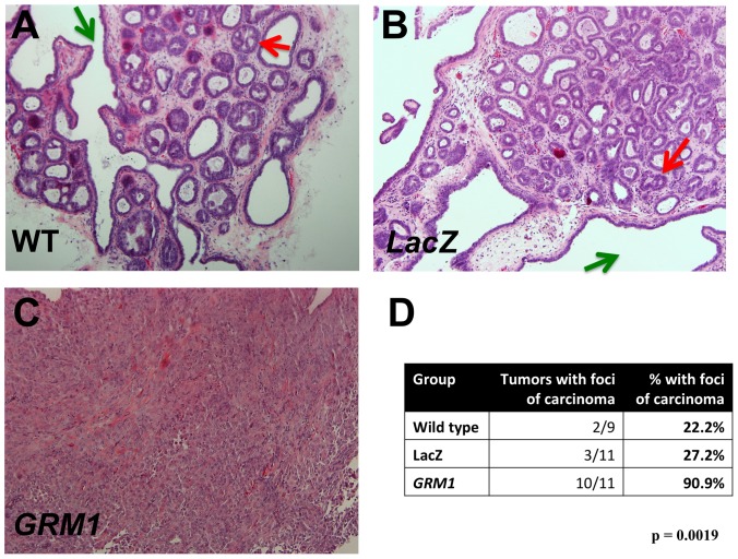
Figure 9. mGluR1 transforms MCF10AT1 cells.
MCF10AT1 cells, wild type or transduced with either LacZ or GRM1, were implanted into both flanks of athymic nude mice and allowed to grow for 8 weeks, after which MCF10AT1 lesions were harvested. A. Representative wild type MCF10AT1 lesion with both papillary enfolding (green arrow) and cribiform foci (red arrow). B. LacZ control MCF10AT1 lesion. The morphology indicates a hyperplastic lesion with both papillary (green arrow) and initial cribiforming present (red arrow). C. mGluR1-overexpressing MCF10AT1 lesion. The morphology indicates invasive cancer. These figures (A through C) are representative of 10 lesions analyzed for each group (magnification: 100×). D. GRM1 overexpression results in malignant transformation of MCF10AT1 cells. Standard H&E sections of tumors were examined by a trained observer blinded to experimental group, and MCF10AT1 xenografts assessed for the presence and number of foci of carcinoma.

