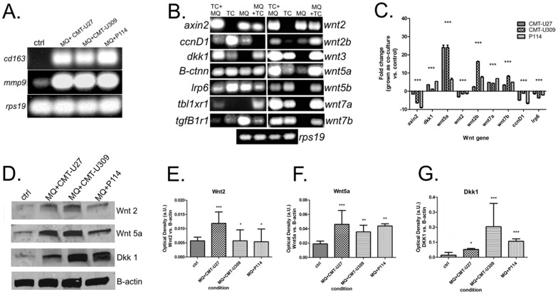
Figure 1. Expression of selected Wnt genes in co-cultured canine mammary tumor cells and macrophages.
Real-time RT-PCR analysis of Wnt genes in co-cultured and monocultured canine mammary neoplastic cells and macrophages. (A) Representative agarose gel electrophoresis of Cd163 and Mmp9 PCR products following real-time SYBR Green amplification in macrophages cultured alone and in co-culture with neoplastic cells. (B) Representative agarose gel electrophoresis of PCR products following real-time SYBR Green amplification (CA, cancer cells grown as monoculture; CA+MQ, co-cultured cancer cells; MQ, mono-cultured macrophages; and MQ+CA, macrophages co-cultured with cancer cells). (C) Fold changes in examined genes in co-cultured macrophages compared with control macrophages. (D) Western blots of cytoplasmic and membrane Wnt proteins from Macrophages grown as monocultures or sorted from co-culture with canine mammary neoplastic cells. The level of examined proteins (E, F, G) was expressed as IOD (Integrated Optical Density) in arbitrary units with the value obtained using the Odyssey Infrared Imaging System (LI-COR Inc., USA). The results are expressed as the mean ±SD. The ANOVA + Tukey post-hoc test were applied (Graph Pad v. 5.0), the values differed significantly (p<0.05) were marked as *, whereas values differed highly significant (p<0.01 or p<0.001) were marked as ** or ***, respectively.

