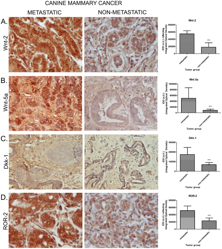
Figure 6. Analysis of Wnt proteins expression in metastatic and non-metastatic canine mammary cancer tissues.
Images showing Wnt-2, Wnt-5a, Dkk-1, and ROR-2 expression in metastatic and non-metastatic canine mammary tumors were obtained using an Olympus BX60 microscope (400×). Tissue sections were treated with specific antibodies and stained using an EnVision kit (Dako, Denmark). Labeled antigens are indicated by brown precipitates. Metastatic tumors show significantly higher expression of all examined antigens compared with non-metastatic tumors. Colorimetric intensities of immunohistochemical-stained antigen spots on 10–20 images were counted using a computer-assisted image analyzer (Olympus Microimage™ Image Analysis version 4.0 software for Windows, USA) and were expressed as mean pixel integrated optical density (IOD). Statistical analysis was performed using Prism version 5.00 software (GraphPad Software, USA). Analysis of variance and Tukey's post-hoc tests were used to identify differences in optical density. Differences were considered significant when *p<0.05, and highly significant when **p≤0.01 or ***p≤0.001.

