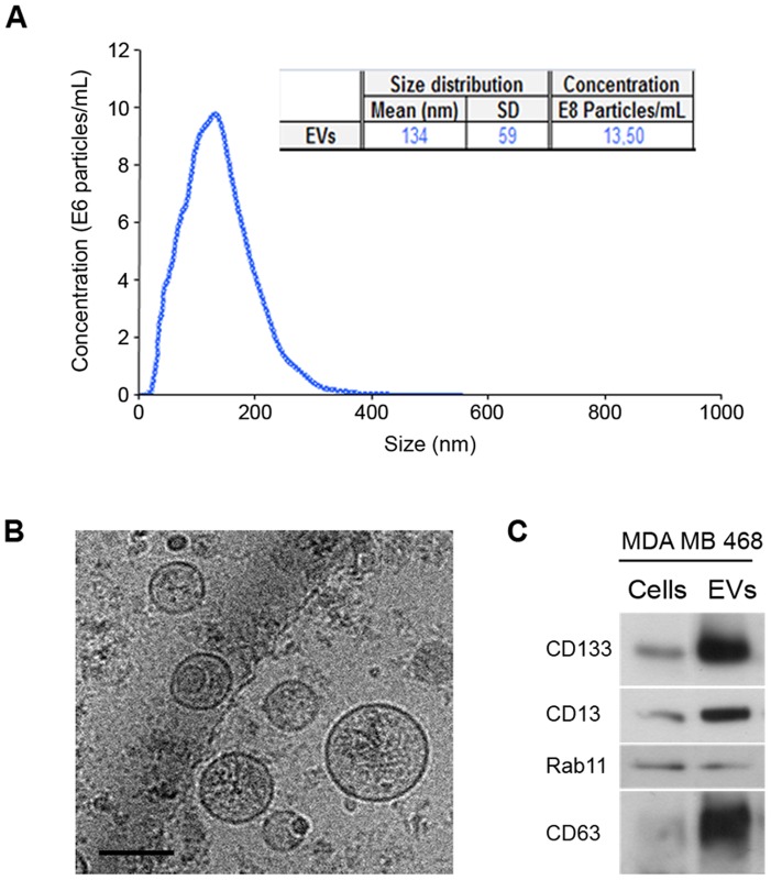
Figure 5. Characterization of EVs secreted by MDA-MB-468 mammospheres.
(A) NTA analysis indicating mean, standard deviation and concentration of the particles present in the preparation. (B) Representative cryo-electron micrographs (Bar, 100 nm). (C) Western blot analysis of protein extracts prepared from cells or from EVs using antibodies against indicated proteins.

